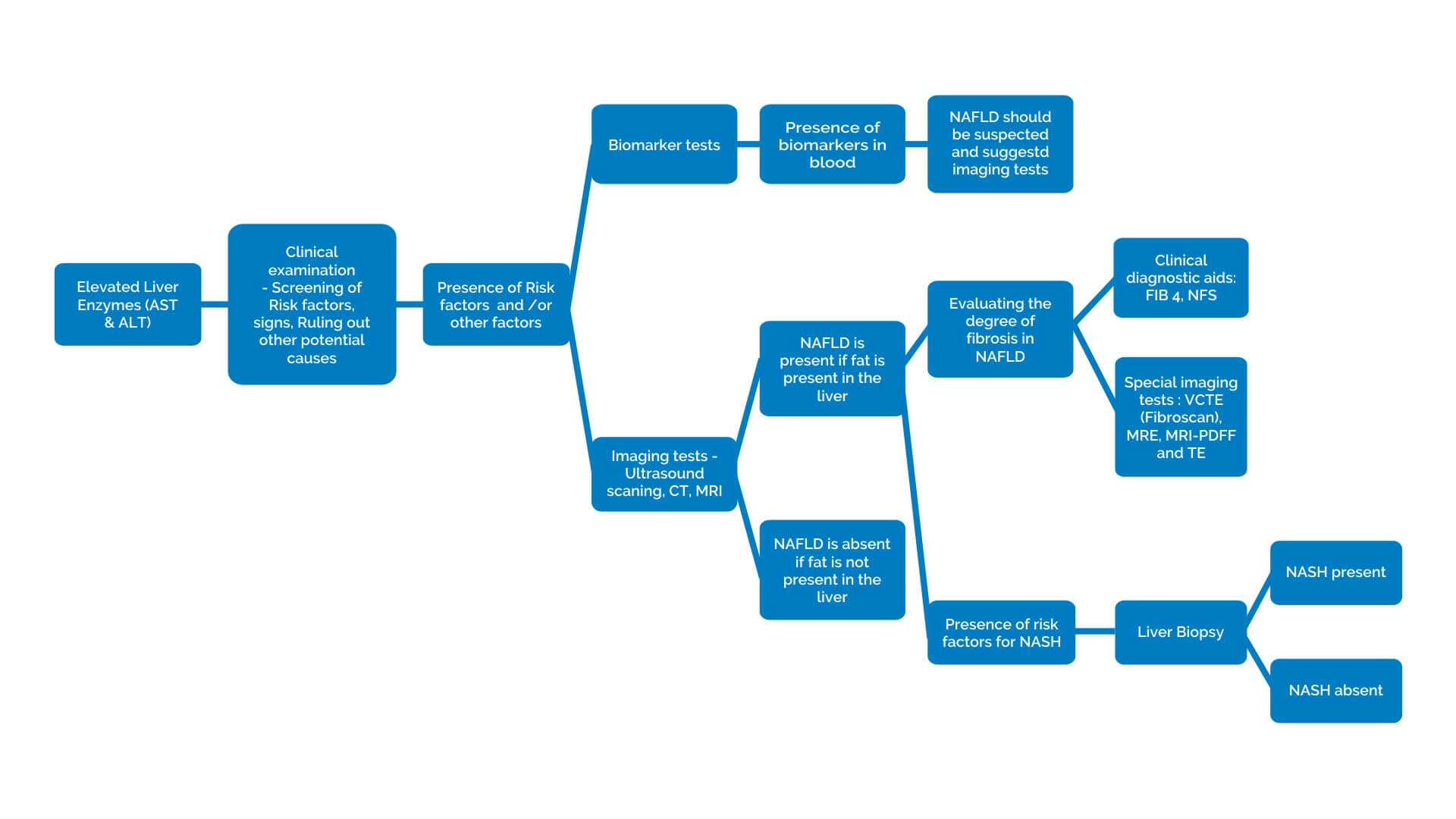
Since both NAFLD and NASH do not show any symptoms they are very often incidentally diagnosed during routine blood tests with elevated liver enzymes or ultrasound scanning done for reasons other than fatty liver. Doctors diagnose both NAFLD and NASH by using various diagnostic tools.
Diagnosis
Diagnosis of NAFLD
How is NAFLD diagnosed?
Diagnostic Tools used in Diagnosing NAFLD and NASH
Clinical examination
Initially your doctor will evaluate you if you have any risk factors of NAFLD and rule out any other potential causes of NAFLD.
Screening for Risk Factors: A thorough medical history of you with questions related to your history of health conditions that are considered as risk factors to develop NAFLD are screened
- Over weight or obesity
- Insulin resistance
- High levels of triglycerides or abnormal levels of cholesterol in blood
- Metabolic syndrome
- Type 2 diabetes
- Insulin resistance
Most importantly the doctor will ask about your alcohol intake to rule out alcoholic liver disease.
Physical examination: During a physical examination, your doctor examines your body to look for signs such as
- An enlarged liver with mild sensitivity
- Insulin resistance like darkening of skin at the neck region,
- Jaundice, where skin and whites of your eyes to turn yellow
- Cirrhosis with water retention in abdomen and legs
Rules out other potential causes of fatty liver: Your doctor will exclude other diagnosed potential causes of fatty liver like
- Other liver disease
- Autoimmune disease
- Viral hepatitis
- Wilson’s disease
- Are on certain drugs
Blood Tests
Serum Biomarker tests
Along with ALT and AST certain other substances and tests also give an idea about liver damage. Biomarker tests are one such type of blood tests that help in identifying certain substances in blood that reflect the presence of particular disease. But it should be remembered that these tests alone may not establish the presence of NAFLD or NASH.

Some of the serum biomarker tests
-
Liver Function Tests (LFT)
-
Lipid Profile (TG,FFA,Cholesterol)
-
Serum autoantibodies
-
Proinflammatory and profibrogenic markers
-
Cytokines
-
Hepatocyte apoptosis markers
Ruling out other potential causes of fatty liver:
While diagnosing NAFLD your doctor will recommend further blood tests that help in ruling out other causes of fatty liver disease (with elevated liver enzymes) like viral hepatitis, autoimmune liver disease, and metabolic or inherited liver disease.
Imaging Studies:
Imaging studies are those studies where the images of the internal organs can be produced giving us a picture of what is inside the body. Imaging studies that are commonly used in the diagnosis of NAFLD are
- Abdominal Ultrasonography
- CT Scan
- MRI Scan
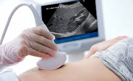
Significance of Imaging studies:
A person will be diagnosed with NAFLD only if there is fat accumulation in liver in imaging studies (ultrasound and CT) with no significant consumption of alcohol, no other competing reasons for fatty Liver and no other co –existing liver diseases.
Challenges of imaging studies:
The imaging studies establish the accumulation of fat in the liver (NAFLD) but cannot differentiate the type of fatty liver, like whether it’s just a simple fatty liver or more serious form like NASH or the degree of fibrosis in the liver.
Diagnosing degree of fibrosis in NAFLD:
To know the whether the degree of fibrosis is advanced, bridging fibrosis (stage 3) or cirrhosis (stage 4) your doctor will use the following
Clinical Decision Aids:
To evaluate the degree of fibrosis or amount of liver tissue that is damaged and replaced with fibrous tissue your doctor will use certain clinical decision tools like FIB-4 index, NFS score that combine the results of your blood tests, with your risk factors for NAFLD.
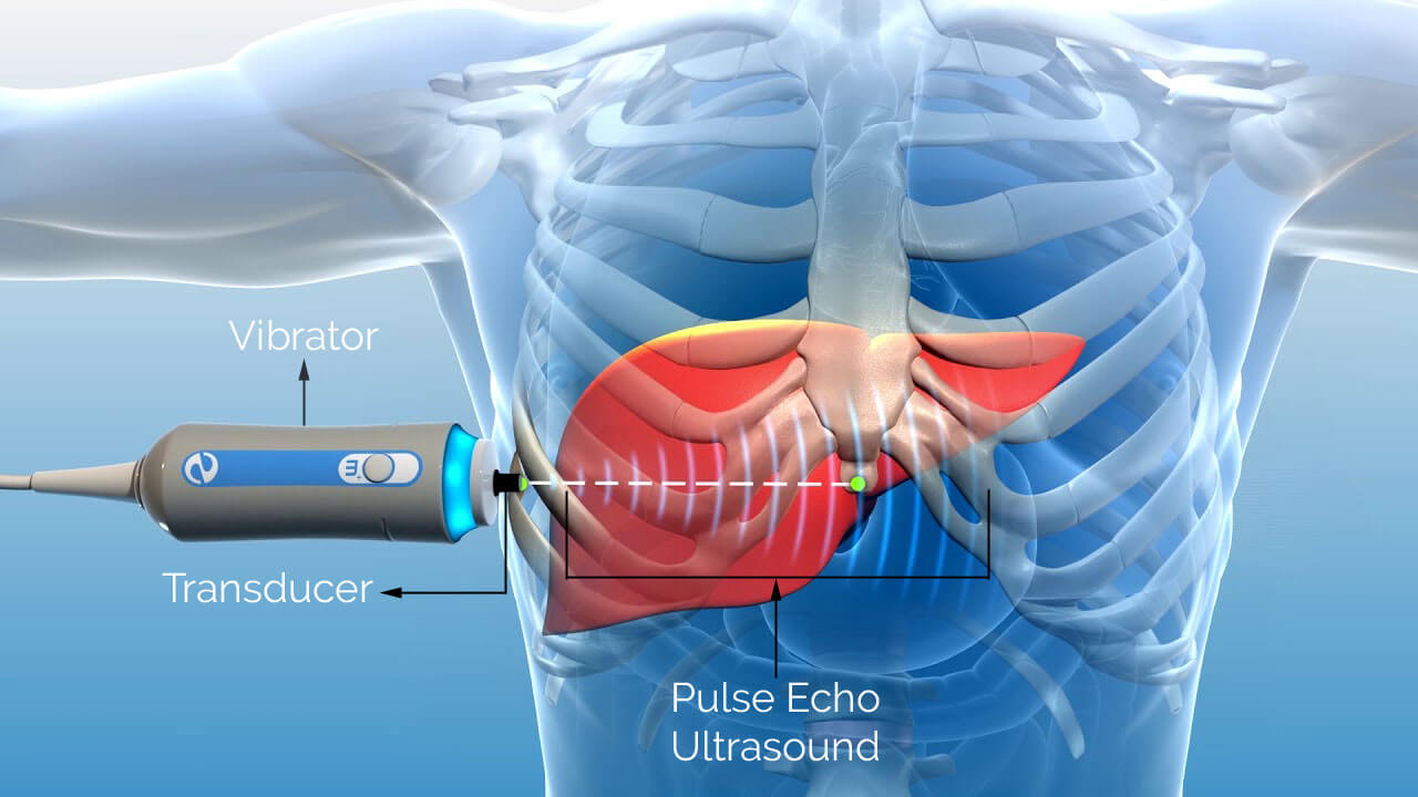
Special scans to measure the stiffness of liver:
The liver becomes stiff due to fibrosis or scar tissue and fat accumulation, normal ultrasound scans can not measure this, but special scans can measure the liver stiffness to ascertain the degree of fibrosis and fat level. Special scans
- VCTE (Fibroscan)
- MRE
- MRI-PDFF
- TE
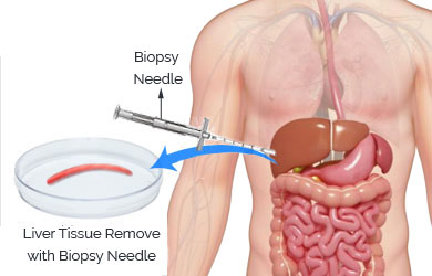
Diagnosis of NASH:
NASH cannot be diagnosed by blood tests and imaging studies. At present, the only reliable way of diagnosing whether a person with fatty liver has NASH is by a liver biopsy.
Liver Biopsy:
Is a procedure where a very small piece of liver is retrieved through a needle and tested under microscope for its contents.
Considerations for Liver Biopsy:
Liver Biopsy is considered only in NAFLD patients with
- Advanced fibrosis
- Are at increased risk of NASH
- Patients who can benefit from diagnosis and treatment
Significance of liver biopsy:
NASH can be diagnosed only by a biopsy and only if the tested piece of liver shows infiltration of the liver with fat along with inflammation and different degrees of scarring.
Who needs to be evaluated for NAFLD?
Your doctor may recommend you for blood tests (liver function tests), and other tests if you have risk factors for NAFLD/NASH and will proceed further depending on the test results.
Diagnosis Algorithm of NAFLD and NASH:
Reference:
- Cleveland, E. , Bandy, A. and VanWagner, L. B. (2018), Diagnostic challenges of nonalcoholic fatty liver disease/nonalcoholic steatohepatitis. Clinical Liver Disease, 11: 98-104. doi:10.1002/cld.716
- Chalasani, N. , Younossi, Z. , Lavine, J. E., Charlton, M. , Cusi, K. , Rinella, M. , Harrison, S. A., Brunt, E. M. and Sanyal, A. J. (2018), The diagnosis and management of nonalcoholic fatty liver disease: Practice guidance from the American Association for the Study of Liver Diseases. Hepatology, 67: 328-357. doi:10.1002/hep.29367
Copyright ©2018 all rights reserved
Designed by Point Blank